1) What Shock Is (Physiology)
Shock = inadequate tissue perfusion. Cells switch to anaerobic metabolism → lactate rises → acidosis impairs enzyme systems. Compensatory responses (tachycardia, vasoconstriction) shunt blood to heart/brain at the expense of skin, gut, and limbs.
2) Major Types You’ll See Outside
- Hypovolemic: blood or fluid loss lowers preload → stroke volume drops.
- Distributive: anaphylaxis/sepsis cause vasodilation and capillary leak → relative hypovolemia.
- Cardiogenic: pump failure (MI, arrhythmia) → poor forward flow despite volume.
- Obstructive (less common): tension pneumothorax, tamponade → mechanical block to filling/flow.
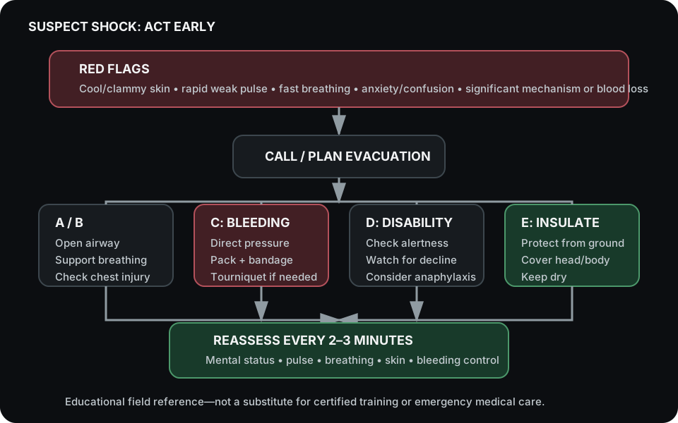
3) Field Recognition (Red Flags)
- Skin: pale, cool, clammy (peripheral vasoconstriction).
- Pulse: rapid & thready; cap refill > 2 s in cool environments is concerning.
- Breathing: fast/shallow (blowing off CO₂ to buffer acidosis).
- Mental status: anxiety → confusion → lethargy (cerebral hypoperfusion).
- Blood loss: obvious bleeding or occult (pelvis/thigh, GI, chest).
4) Immediate Actions (ABCDE, Stop the Bleed)
- A/B: Airway & Breathing — open airway; high-flow supplemental O₂ if available; watch for chest wounds.
- C: Circulation — control hemorrhage first: direct pressure → hemostatic packing → pressure bandage → tourniquet (mark time).
- D: Disability — check level of consciousness; look for anaphylaxis signs (rash, wheeze, swelling).
- E: Exposure/Environment — prevent heat loss; use blanket/insulation. Hypothermia worsens coagulopathy.
Positioning: Position the patient according to airway, breathing, injuries, comfort, current training, and local protocol. Avoid a universal positioning rule; different causes of shock and associated injuries require different decisions.
5) Biological Rationale for Key Interventions
- Tourniquets: Rapid arterial occlusion prevents ongoing volume loss; pain is expected—do not loosen for comfort.
- Hemostatic Gauze: Kaolin activates intrinsic pathway; chitosan provides a positively charged matrix to aggregate RBCs—both accelerate clot formation.
- Heat Preservation: Coagulation enzymes work poorly when cold; shivering raises metabolic demand—insulate early.
- Epinephrine (Anaphylaxis): Alpha/beta agonism reverses vasodilation/bronchospasm; auto-injectors if trained and prescribed.
6) Monitoring & Evac
- Reassess every 2–3 minutes: mental status, pulse quality, breathing, skin temperature, bleeding control.
- Document times: tourniquet application, meds given, changes in status.
- Evacuate early—shock can look “okay” then crash as compensation fails.
7) Field Drill (10–12 minutes)
- Team of 3: Airway/Breathing lead, Bleeding control, Recorder.
- Simulate arterial bleed (training prop). Apply pressure → pack wound (3 minutes) → pressure bandage → tourniquet if needed.
- Recorder logs vitals every 2 minutes and times interventions.
- Wrap patient to prevent heat loss; reassess; prep for evac.
Equipment research is separate
For commercial supplier notes and product comparisons, continue to Shop Gear. Equipment does not replace certified training, scope, protocol, or medical direction.